Prof.Ding's Research Group
Department of Biomaterials and Tissue Engineering, College of Life Sciences
State Key Laboratory of Medicinal Chemical Biology
State Key Laboratory of Medicinal Chemical Biology
Organic
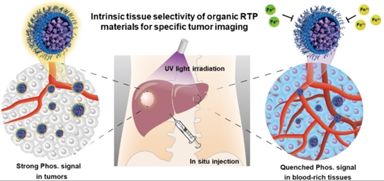
phosphorescent materials are excellent candidates for use in tumor imaging.
However, a systematic comparison of the effects of the intensity, lifetime, and
wavelength of phosphorescent emissions on bioimaging performance has not yet
been undertaken. In addition, there have been few reports on organic
phosphorescent materials that specifically distinguish tumors from normal
tissues. This study addresses these gaps and reveals that longer lifetimes
effectively increase the signal intensity, whereas longer wavelengths enhance
the penetration depth. Conversely, a strong emission intensity with a short
lifetime does not necessarily yield robust imaging signals. Building upon these
findings, an organo-phosphorescent material with a lifetime of 0.94 s
was designed for tumor imaging. Remarkably, the phosphorescent signals of
various organic nanoparticles are nearly extinguished in blood-rich organs
because of the quenching effect of iron ions. Moreover, for the first time, we
demonstrated that iron ions universally quench the phosphorescence of organic
room-temperature phosphorescent materials, which is an inherent property of
such substances. Leveraging this property, both the normal liver and hepatitis tissues
exhibit negligible phosphorescent signals, whereas liver tumors display intense
phosphorescence. Therefore, phosphorescent materials, unlike chemiluminescent
or fluorescent materials, can exploit this unique inherent property to
selectively distinguish liver tumor tissues from normal tissues without
additional modifications or treatments.

 津公网安备 12010402001780号